Left Hepatectomy [Case 9]
Introduction
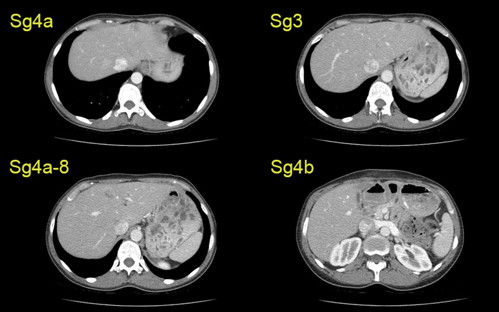
A 43-year old woman with breast cancer has synchronous liver metastases in segments 3, 4a, 8/4a and 4b. After undergoing ablation and radiation, the liver metastases recurred. Over two years, she responded to chemotherapy and hormone therapy. A left hepatectomy was recommended.
Left Hepatectomy [Case 9]
Imaging Analysis
Look through the patient's CT scan to identify any areas of concern for the necessary procedure.
Left Hepatectomy [Case 9]
Surgeon's Impressions
Watch the following videos of the surgeons preoperative plans for the case. Compare their findings with your own analysis.
Summary of surgeons’ findings and slowing down moments:
Operating surgeon
Dr. Gallinger's critical imaging findings:
- Sg6-7 posterior right hepatic duct
- Tumor proximity to middle hepatic vein
Consulting surgeon
Dr. Moulton's critical imaging findings:
- Tumor proximity to middle hepatic vein
- Left portal vein anatomy
- Hepatic vein branches
- Lesion in Sg4b
- Gallbladder thickened
- Determining liver transection plane
Watch the following videos from the operation to see some of the key moments in the case.
Watch the following video for the operating surgeon's summary of the case.
The issues that the surgeons collectively identified preoperatively:
- Sg6-7 posterior hepatic duct
- Tumor proximity to middle hepatic vein
- Left portal vein anatomy
- Hepatic vein branches
- Lesion in Sg4b
- Determining liver transection plane
- Gallbladder thickened