Left Lateral Sectionectomy [Case 6]
Introduction
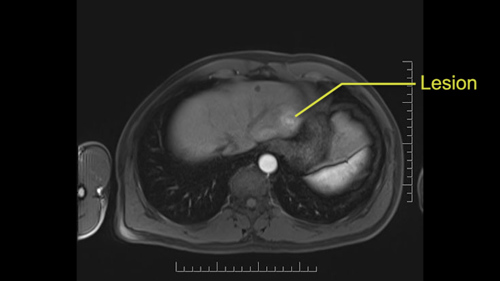
A 56-year old man was diagnosed with uveal melanoma and underwent enucleation four years ago. He was recently found with metastatic melanoma, on ultrasound and CT, with a metastatic lesion in Segment 2-3 of the liver.
Our plan for him is to do a laparoscopic left lateral sectionectomy.
Left Lateral Sectionectomy [Case 6]
Imaging Analysis
Look through the patient's CT scan to identify any areas of concern for the necessary procedure.
Axial Venous

Insert Image Sequence here
Left Lateral Sectionectomy [Case 6]
Surgeon's Impressions
OR Footage
Summary of surgeons’ findings and slowing down moments:
Operating surgeon
-
Dr. Cleary's procedure-specific moments:
- Dissection of biliary plate
- Dissection of left hepatic vein
-
Patient imaging findings:
- Indeterminate lesion in Segment 4
- Replaced left hepatic artery
- Left hepatic vein well-demarcated
Consulting surgeon
-
Dr. Moulton's imaging findings:
- Accessory left hepatic artery
- Segment 4 branch of left hepatic vein
- Possible left hepatectomy for Sg4 lesion
-
Procedure-specific moments:
- Dissection of left hepatic vein
- Tumor proximity to left portal vein
Watch the following videos from the operation to see some of the key moments in the case.
Watch the following video for the operating surgeon's summary of the case.
Key points from this case
Indeterminate lesions: include intraoperative ultrasound and be prepared to change the operative plan if a larger procedure is required. But be mindful of new procedure-specific slowing down moments and keep in mind that the exposure and ports may be different from usual.
The issues that the surgeons collectively identified preoperatively:
- Indeterminate lesion in Segment 4
- Replaced left (accessory) hepatic artery
- Segment 4 branch of left hepatic vein
The issues were demonstrated in the intraoperative videos. On the pre-operative MRI, there was an indeterminate lesion in segment 4 and when we looked at that on ultrasound, we felt that it was bigger than it was on MRI and therefore suspicious for another lesion. So we ended up doing, instead of a left lateral sectionectomy which is segment 2-3, we did a left lobectomy which is segments 2, 3, 4a, 4b. It’s a slightly harder operation.
It requires a portal dissection and the first slowing down moment was really on the ultrasound. Another slowing down moment was on the artery, on the left portal vein, on the left bile duct and then obviously on the left hepatic vein, and then obviously the orientation is a little bit different but we then had a cholecystectomy, you know, which is usually most of the time just a very routine part of the operation but I think when the exposure is different, your ports are different, you have to spend a little bit more time and be a little bit more careful.
Anyways, so those were our moments. There were no real complications during the operation; everything seemed to be going relatively smoothly so from that perspective it was just a bigger operation than normal, than we expected, and a little more complex but there was nothing during the operation that was problematic.