Distal pancreatectomy with splenectomy [Case 5]
Introduction
Clinical History
A 58-year old woman presented with abdominal discomfort. CT imaging demonstrated a cystic lesion in the tail of her pancreas, as well as splenic vein thrombosis. A distal pancreatectomy with splenectomy was recommended.
Distal pancreatectomy with splenectomy [Case 5]
Imaging Analysis
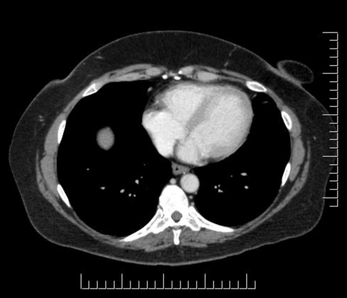
Look through the patient's CT scan to identify any areas of concern for the necessary procedure.
Axial Venous

Distal pancreatectomy with splenectomy [Case 5]
Surgeon's Impressions
Summary of surgeons' findings and slowing down moments:
Operating surgeon: Dr. Moulton
- Procedure-specific moments:
- Tumor proximity to splenic artery
- Tumor proximity to splenic vein
- Tumor proximity to portal vein
- Critical imaging findings:
- Retroperitoneal margin
- Left-sided portal hypertension
- Replaced right hepatic artery
Consulting surgeon: Dr. Cleary
- Procedure-specific moments:
- Medial mobilization and dissection of pancreas
- Transecting neck of pancreas
- Splenic dissection
- Critical imaging findings:
- Left-sided portal hypertension
- Replaced hepatic arteries
- Tumor proximity to splenic vein, PV confluence
- Tumor proximity to splenic artery, celiac axis
- Retroperitoneal dissection
Watch the following videos from the operation to see some of the key moments in the case.
Watch the following video for the operating surgeon's summary of the case.
Key points from this case
- Retroperitoneal dissection: anticipate a more challenging dissection in the retroperitoneal margin when there is inflammation, and be aware of varices and collaterals in cases of left-sided portal hypertension.
The issues that the surgeons collectively identified preoperatively:
- Left-sided portal hypertension
- Retroperitoneal dissection
- Tumor proximity to blood vessels (splenic artery, splenic vein)
- Replaced right hepatic artery
The first 3 issues were demonstrated in the intraoperative videos.
We just completed the distal pancreatectomy and splenectomy. It went pretty much as planned. Quite challenging because of the depth of the patient and lots of adipose tissue.
My normal approach is: to take down the splenic flexure come along the inferior border of the pancreas to the neck, past the lesion, open up the retroperitoneal plane, which we did. And we actually did find that it was quite challenging and sticky as we expected on the CT scan. Then we divided the splenic artery, which was a little bit difficult, stuck on to the superior border of the pancreas so we had to go more proximal than I thought to take that.
Then the splenic vein, we took that in the usual fashion with a stapler, and then the neck of the pancreas with a stapler. Then we continued along the superior border and the retroperitoneal surface of the pancreas, mobilized the spleen and it was out.
So, all-in-all, no real surprises. The left-sided portal hypertension and varices were a challenge but not necessarily with a big ligasure. I think it would have been a challenging laparoscopic case.