Extended Left Hepatectomy [Case 3]
Introduction
A 73-year old man presented with jaundice. CT imaging showed a presumed cholangiocarcinoma at the bile duct bifurcation (Klatskin tumor).
An extended left hepatectomy was recommended.
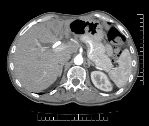
Imaging Analysis
Look through the patient's CT scan to identify any areas of concern for the necessary procedure.
Axial Arterial

Extended Left Hepatectomy [Case 3]
Surgeon's Impressions
Watch the following videos of the surgeons preoperative plans for the case. Compare their findings with your own analysis.
Patient-specific findings
- Portal vein involvement: LPV stricture, RPV free
- Arterial involvement: LHA involved; RHA from SMA, RHA clear of tumor
- Left hemihepatectomy with caudate resection
- Frozen section of both bile duct margins
Summary of surgeons' findings and slowing down moments:
Operating surgeon
- Dr. McGilvray's imaging findings:
- Tumor proximity to portal vein bifurcation
- Tumor proximity to bile duct bifurcation
- Plane of liver transection
Consulting surgeon
-
Dr. Moulton's procedure-specific moments:
- Caudate inflow/outflow
- Left portal vein
- Left hepatic artery
- Right hepatic artery
- Left hepatic vein
-
Patient imaging findings:
- Replaced right hepatic artery
- Tumor proximity to middle hepatic vein
- Caudate inflow/outflow
- Tumor proximity to left portal vein
Watch the following videos from the operation to see some of the key moments in the case
Watch the following video for the operating surgeon's summary of the case.
Key points from this case
In a Klatskin tumor, evaluate the portal vein and the distal aspects of the hepatic artery for oncologic margins. If you do determine that you have to resect and reconstruct any vascular structures, its better to commit to doing that sooner rather than later, so that you can set it up and be ready for it.
The issues that the surgeons collectively identified preoperatively:
- Replaced right hepatic artery
- Tumor proximity to portal vein bifurcation
- Tumor proximity to bile duct bifurcation
- Determining the plane of liver transection
The four issues were demonstrated in the intraoperative videos. It was an extended left hepatectomy. This was a reasonably straightforward left-dominant Klatskin. My strong bias in cases like this is to take any vascular structures that are potentially compromised so as to compromise the oncologic margin. But in this case, all of the vessels fell away from the tumor specimen quite readily.
So one question that comes up from a case like this is when you would commit to doing a portal vein resection and reconstruction, or indeed an arterial resection and reconstruction in the context of a Klatskin. One of the tricks of doing a Klatskin is you tend to take the bile duct complex early so that you can evaluate the portal vein to some extent so that you can evaluate the distal aspects of the hepatic artery.