Vasculobiliary Injury [M28]
Introduction
Clinical History
The patient is a 22-year old woman who initially presented in the emergency department with acute cholecystitis and signs of choledocholithiasis. She underwent an open cholecystectomy with bile duct exploration. According to the surgical report, there was inflammation around the gallbladder that made exposure and dissection difficult. Some bleeding was encountered in the hepatoduodenal pedicle which required stitches to repair.
On post-operative Day 1, the patient required monitoring in the intensive care unit due to decrease in consciousness. The patient's CT scan revealed portal vein thrombosis, right liver ischemia and ascites. With deterioration of consciousness and Grade 2 encephalopathy, on post-operative Day 4 she was transferred to an HPB centre.
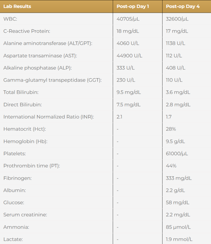
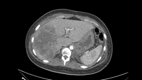
Imaging revealed a right liver infarction, right poral vein thrombosis extending to the main portal vein. Both the right and left hepatic arteries were absent but normal arterial flow was identified through an accessory branch off the left gastric artery. The consensus diagnosis is a vasculobiliary injury.
Vasculobiliary Injury [M28]
Imaging Analysis
Look through the patient's CT scan to identify any areas of concern for the necessary procedure.
Discussions
Watch the following videos from the operation to see some of the key moments in the case.
Watch the following videos for the operating surgeon's summary of the case.
Key points from this case
-
Presence of an accessory artery to the left liver helped to preserve liver function to give us the option for resection.
-
The deteriorating condition of the patient and available options weigh in on the timing of choosing surgical treatment.
The patient went very well. This is the CT scan done a few days after our surgery. Because the patient had some fever and she had some sub-phrenic collection. But the interesting thing to see on the CT scan is how the left sector of the liver was re-perfused with a good portal and arterial flow. There’s some free collection in the right hepatic liver but you can see here how the portal vein is flowing very well. This is the portal vein with a good flow coming from the SMV. You can see here. All of this were blocked by thrombus as we saw in the first CT and now its totally open and you see how this is the Rex process of the portal vein inside the liver, the branches to Sg2 and 3 and also to Sg4. The other important thing to see here is the artery, here the left artery is flowing very well and is also patent. Remember that this is not the proper artery, this is an accessory artery coming from the stomach but it was enough to perfuse all the liver.
So in summary, this was a successful treatment of a very young woman with a complex vasculobiiliary injury after an open cholecystectomy plus bile duct exploration for choledocholithiasis. And this procedure was very important to avoid an emergency liver transplant for the patient and for the the reoperation for the liver dysfunction, portal vein reconstruction. So we were able to identify the Rex process and do a Rex shunt from the SMV.
Again the presence of the accessory left hepatic artery from the left gastric artery, probably favored that the patient not remain total anhepatic after the original procedure of open cholecystectomy and she was able to maintain the arterial perfusion in the lateral sector of the liver with a minimum of liver function which allowed to survive this patient and give some time to do the final repair with this shunt from the SMV. Thank you very much.