Laparoscopic Sg5-6 Resection [Case 27]
Introduction
The patient is a 58-year old gentleman with chronic Hepatitis B. He was found to have a single hypervascular 7.6 x 7.1 cm hepatoma lesion in Segment 5-6. On imaging, there is no evidence of ascites or portal hypertension. His platelet count and bilirubin are normal. The liver has a small degree of fibrosis from cirrhosis. Our plan for him is to do a Segment 5 and 6 resection.
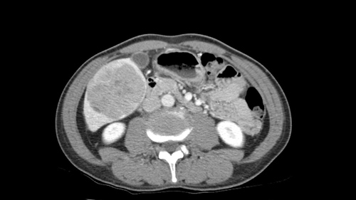
Laparoscopic Sg5-6 Resection [Case 27]
Imaging Analysis
Look through the patient's CT scan to identify any areas of concern for the necessary procedure.
Watch the following videos of the surgeons preoperative plans for the case. Compare their findings with your own analysis.
Summary of surgeons’ findings and slowing down moments:
Dr. Sapisochin's patient-specific planning
- Fibrotic liver, normal function
- No portal hypertension or varices
- Right anterior pedicle Sg 5 branch
- Preserve Sg 7,8
- Dissect right posterior pedicle: Sg 6 branch
- Intraoperative ultrasound: identify transection plane
- Pringle maneuver
Consulting surgeon
Dr. Jayaraman's patient-specific planning
- Normal liver, no varices or portal collaterals
- PV branching; ligate Sg 6 extrahepatic, Sg 5 intrahepatic
- Open resection
- Intraoperative ultrasound: Sg 5-8 margins, outflow
- Preserve Sg 7,8
- Preserve RPV, outflow
- Careful handling
Watch the following videos from the operation to see some of the key moments in the case.
Watch the following video for the operating surgeon's summary of the case.
Key points from this case
- Take care in identifying the anterior and posterior inflow and outflow branches to preserve the remnant liver.
- Applying a Pringle maneuver to reduce blood loss also keeps the transection plane dry for clear dissection and identification of structures.
- When deciding between a laparoscopic or open approach to a Sg5-6 resection, consider your team’s experience with handling a large tumor.
The issues that the surgeons collectively identified preoperatively:
- Normal liver
- Sg6 right posterior pedicle
- Preserve Sg7-8 inflow and outlfow
- Plan for Pringle maneuver
- Ultrasound to confirm margins
- Sg 5 right anterior pedicle branch
The issues were demonstrated in the intraoperative videos. You know this was a patient with a large hepatocellular carcinoma on a fibrotic probably non cirrhotic liver. And the key points here are you know we just need to make sure we preserve all the structures going to the anterior sector and obviously to Segment 7 to be able to safely do a 5-6 resection. As you can see, for me it’s very important to have an intermittent Pringle maneuver and always doing Pringle maneuver as we go and using an ultrasonic device so we can really skeletonize all the structures. And being able to see at any time being what are the pedicles etc. Here we you know identify very well the pedicle going to 5 and to 6. We’re able to take those two pedicles and then to preserve all the right hepatic vein drainage to the segment 7 and some of obviously 5 and 8. And I think those are basically the key points in this case.