Laparoscopic Segment 6 Resection [Case 26]
Introduction
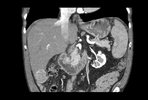
The patient is a 66-year old man with hepatocellular carcinoma and liver cirrhosis. He was found to have a singular 3-cm lesion in Segment 6. On imaging, there is evidence of ascites, mild portal hypertension with small varices and an enlarged spleen. His platelet count and bilirubin are normal. The patient has advanced heart disease so transplant was not recommended. Our plan for him is to do a laparoscopic Segment 6 resection.
Laparoscopic Segment 6 Resection [Case 26]
Imaging Analysis
Look through the patient's CT scan to identify any areas of concern for the necessary procedure.
Surgeon's Impressions
Watch the following videos of the surgeons preoperative plans for the case. Compare their findings with your own analysis.
OR Footage
Summary of surgeons’ findings and slowing down moments:
Operating surgeon
Dr. Sapisochin's patient-specific moments:
- Cirrhotic liver, mild portal hypertension, some ascites
- Intermittent Pringle maneuver
- Careful dissection with CUSA, skeletonize pedicles and structures
- Sg6 pedicle bifurcation
- RHV branch in Sg7
- Slight mobilization of right lobe
- Ultrasound to confirm margins
Consulting surgeon
Dr. Jayaraman's patient imaging findings:
- Favorable location for laparoscopic resection
- Cirrhotic liver
- Ascites
- Collaterals (varices): careful mobilization
Procedure moments:
- Intraoperative ultrasound: exclude other lesions; ensure adequate margins
- Patient positioning for greater exposure, access to porta hepatis
- Pringle maneuvre; careful opening of pars flaccida
Watch the following videos from the operation to see some of the key moments in the case.
Watch the following video for the operating surgeon's summary of the case.
Key points from this case
- Be prepared in setting up a Pringle maneuver in patients with cirrhosis as minimizing blood loss is a concern even for small segmental resections.
- Keep in mind that transecting through a cirrhotic liver parenchyma may be more challenging.
- Be sure to carefully identify all pedicles and vascular structures.
The issues that the surgeons collectively identified preoperatively:
- Cirrhotic liver
- Ascites
- Portal hypertension, varices
- Plan for Pringle maneuver
- Ultrasound to confirm margins
- Encountering Sg6 pedicle, RHV branch
The issues were demonstrated in the intraoperative videos. So as anticipated, once we entered the abdomen, we can obviously see a really cirrhotic liver. There was a small amount of ascites as also anticipated. The tumor was hanging on Segment 6. There were some adhesions to the gallbladder that were easy to take down. We were able to very easily access the back of the porta to put our Pringle. With the help of the ultrasound machine we can identify the tumor and make sure we were transecting with a safe margin and actually transecting and identifying the branches of the Segment 6 pedicle and completing what would be probably mostly a Segment 6 resection.
The transection was a bit more challenging than expected because the liver was very very stiff. In cirrhotic patients the liver is very difficult to open up as a book and the CUSA device obviously even though it's a very efficient machine, it didn’t transect as well as on a healthy liver. However we were able to identify all the pedicles, dissect them and take them with Hem-o-loks. As we transected the liver, the liver started to open up and we were able to do a safe transection.
We had a discussion about leaving a drain and given that he already had ascites and it’s likely that he’s going to develop more ascites, we decided to leave a drain in the area of the transection. Before removing the specimen, we actually decreased quite a bit the pneumoperitoneum to make sure there was no bleeding and once we were happy with hemostasis, we finished the case. It was more or less as we anticipated it would be.