Laparoscopic Left Lateral Liver Resection [Case 25]
Introduction
This is a 74 year old female patient who presented with abdominal pain. On imaging, a lesion in Segment 2-3 of the liver was identified as being suspicious for malignancy. CT and MRI Primovist imaging were done, which shows capsular retraction, hepatic steatosis and some perfusional change surrounding the lesion. The lesion demonstrated interval increase in size and at last imaging measures 2.7 x 1.6cm in size. Her CA19-9 was 36.
A second lesion in Segment 5/7 is stable and after an additional ultrasound found no signs of vascularity, this was unlikely to be a neoplasm. She has had a previous laparoscopic cholecystectomy which could account for some mild biliary dilatation.
Our plan is to do a laparoscopic left lateral sectionectomy. Notably, the patient has requested not to receive any blood products.
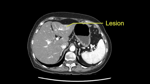
Laparoscopic Left Lateral Liver Resection [Case 25]
Imaging Analysis
Look through the patient's CT scan to identify any areas of concern for the necessary procedure.
Left Hepatectomy [case 1]
Surgeon's Impressions
OR Footage
Summary of surgeons' findings and slowing down moments:
Operating surgeon, Dr. Moulton
-
Procedure-specific moments:
- Port positioning
- LPV inflow to Sg2,3
- Proximity of LHV
-
Patient-specific moments:
- Transection margin at LHV branches run parallel
- Pringle maneuvre: previous cholecystectomy, Foramen of Winslow
- Avoid portal dissection
Consulting surgeon, Dr. Sapisochin
-
Critical moments:
- Consider Sg2 resection
- Intermittent Pringle
- Skeletonize structures with control, CUSA
- Identify Sg2, Sg3 pedicles
- Ultrasound to confirm tumor location, relation to LHV
Watch the following videos from the operation to see some of the key moments in the case.
Post-op Debrief
Watch the following videos for the operating surgeon's summary of the case.
Key points from this case
- During mobilization in a left lateral sectionectomy, be careful of phreniv vein coming into the left hepatic vein. Avoid dissecting too medially.
- When encountering large veins during transection, always check the anatomy and confirm on patient imaging before dividing.
The issues that the surgeons collectively identified preoperatively:
- Transection margin and LHV branches
- LPV inflow to Segment 2,3
- Pringle maneuvre
So the slowing down moments that we had in this case prior were port placement. I think we did struggle actually with the view to the liver. During the surgery I didn’t anticipate that the left lobe of the liver is actually kind of more on the patient’s right side than the patient’s left side. She has a very deep liver and you can see here that her liver almost ends in the midline so she has this big floppy segment of liver inferiorly so just trying to get that exposed with the proper angle with our stapler and initial dissection was a little challenge but it was okay we figured it out. We actually just moved the camera over to the patient’s right hand side or right hand sided port which was also 5-12.
The second thing I think that we did was actually not so challenging, it went pretty straightforwardly, was the Pringle maneuvre. I usually approach the foramen of Winslow from the patient’s right hand side with a 5-cm port. There, I introduce the umbilical tape and I usually bring it through and across to the other side and then bring it up through the port, take the port out and replace it with a chest tube to perform the Pringle maneuvre. In this particular case because of the anatomy and because we were and you know the patient’s liver was kind of so far to her right, we actually approached the porta from the left hand side this time and that seemed to work actually quite well as well. Luckily there was a space behind the porta so her previous cholecystectomy did not interfere. The other thing is that when we do a Pringle maneuvre, we put it on for 10 minutes at a time and then leave it off for 5 minutes and I make sure that the communication with the nursing staff is kind of very clear. I think it is important that they recognize when I take the Pringle off and when I put it on and that we confirm back and forth. There was an instance where they thought that I heard them take the Pringle maneuvre off and I think 10 minutes later we were kind of engrossed in our dissection and they thought that we had taken it off and we hadn’t so I think since then even though thats not a big deal, it taught me that we need to kind of confirm back and forth. So communication is very important.
The stapling of the inflow actually went fine. The inflow were exactly where we thought they were. And the slowing down moment during surgery which I had anticipated prior actually also became a slowing down moment intraoperatively at the hepatic vein. You can see here that the hepatic vein is here very close to the inflow but about here we saw that this vein was actually coming in also. And so we just went over and checked on the images kind of during surgery just to make sure we knew exactly what we were dividing, it was a fairly large vein. I think we’re coming across it here again, after we had taken it maybe actually in the liver parenchyma here.
There was a part at the beginning of the procedure where we dissected along the left triangular ligament probably too far medially as you could see the phrenic vein coming into the left hepatic vein or the cava and we actually got into the phrenic vein as it entered the cava and you know there was a little bit of bleeding, there wasn’t much, we actually turned up the pressures. That’s one maneuvre to decrease the bleeding and then we put staples on and it was fine.
Other than that, we left some Surgicel on the cut edge of the liver surface. There was no bleeding at the conclusion of the operation. We put the specimen in a large bag and brought it out through the umbilical incision. Usually I just extend the umbilical incision a little bit through the umbilicus and a little bit below and that provides a really nice kind of almost non-noticeable scar at the umbilicus.