Total Pancreatectomy [Case 24]
Introduction
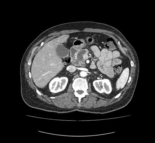
This is a 67 year old man who initially presented with weight loss. His initial ultrasound and subsequent CT scan demonstrated a very large pancreatic duct and a cyst in the pancreas. He had an endoscopic ultrasound which showed thickened septi in the pancreatic cyst. A fine needle aspirate was done and confirmed for mucinous cyst, suggesting IPMN (intraductal papillary mucinous neoplasm).
In addition, an ERCP revealed a fish mouth papilla with mucus coming out of it. His main pancreatic duct is dilated (1.7cm) in the head. The complex cystic mass measures approximately 3.4 x 3.1cm on axial imaging. The pancreatic body appears to have additional small cysts or side branches. There is no suspected vascular invasion, and no biliary dilatation.
Our plan is to prepare for a total pancreatectomy. He has also been consented for a splenectomy just in case.
Total Pancreatectomy [Case 24]
Imaging Analysis
Look through the patient's CT scan to identify any areas of concern for the necessary procedure.
Watch the following videos of the surgeons preoperative plans for the case. Compare their findings with your own analysis.
OR Footage
Summary of surgeons' findings and slowing down moments:
Operating surgeon, Dr. Moulton
-
Patient imaging findings :
- Possible splenectomy: artery, vein preservation
- Proximity of cyst to SMA, renal vein
-
Slowing down moments :
- Mobilize splenic flexure
- Possible splenectomy: Short gastric vessels
- Inferior pancreas: IMV, splenic vein, duodenum
- Kocherization
- Superior pancreas dissection: hepatic artery
- PV-SMV at pancreas neck
- Rotate pancreas
- Uncinate dissection: SMA, IPDAs
Consulting surgeon, Dr. McGilvray
-
Patient imaging findings:
- No mass, no biliary dilatation
- Some soft tissue around SMA, likely a cyst
- Aortic disease, thick walled
- No venous involvement
- Kocherize, assess vessels and rule out mass
-
Slowing down moments:
- Dissect base of pancreas before SMV
- Adequate portal dissection to safely identify portal vein
- Lift pancreas up from retroperitoneum before ligating splenic vein
- Ensure adequate exposure along arteries
- If exposing SMA, locate the root first to ensure control
Watch the following videos from the operation to see some of the key moments in the case.
Watch the following video for the operating surgeon's summary of the case.
Key points from this case:
Hepatic arterial anatomy can be variable. Always confirm the presence of a left and right pulse prior to taking GDA. Also ensure that there is no retrograde flow from the SMA by clamping (bulldog) the GDA and ensuring adequate flow.
The main issues that the surgeons collectively identified preoperatively:
- Soft tissue around SMA, renal vein
- Possible splenectomy
- Kocherization to assess vasculature
- Superior pancreas border
- Inferior pancreas border
- Mobilizing pancreas from retroperitoneum
- Uncinate dissection
So everything went well. We did take the spleen, mostly because as we were actually dividing the short gastrics and mobilizing the distal part of the pancreas we got some bleeding from the capsule of the spleen. So I think I may have said it before, I have a lower threshold for taking this spleen with splenic bleeding so we ended up taking the spleen.
There was a couple of slowing down moments. One, I think the patient was deeper than I had expected and perhaps could have extended the wound at some point but I felt like I was always struggling. Whether the patient was not fully relaxed, the wound was too small or my retractors weren't quite right. I just felt like it wasn't. I was kind of in that groove that I like to be in and everything seemed to be a bit of a struggle.
The second thing was as we were dividing the gastroduodenal artery, we divided the gastroduodenal artery. But then as we were dissecting along the right side of the common hepatic artery there was another artery that was coming down parallel to the gastroduodenal artery. I thought at the time maybe this was two gastroduodenal arteries but you know if you look back on the film you can actually see that there's a smaller branch that is coming beside it as a second gastroduodenal artery. And so I was happy taking that we were taking that and then everything after that was OK.
So in his particular case the bile duct was actually quite small as expected. There was no obstruction. So the bile duct was thin and small in diameter. And so I think it is important to kind of pay particular attention to that when you're doing the reconstruction, especially in this case the pathology is benign and likely he will live for a long time. So I think it's important too to pay particular attention to the bile duct. In this case we used a double ended 6-0 PDS. It was all fine and I think everything went well and that's it.