Extended Left Hepatectomy [Case 20]
Introduction
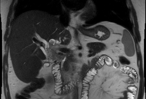
This is a 45-year old man who presented with transient painless jaundice. Imaging showed a large intraductal biliary neoplasm with Klatskin-type tumor that extended down toward his pancreas. An extended left hepatectomy with caudate and extrahepatic bile duct excision and hepaticojejunostomy reconstruction was recommended.
Extended Left Hepatectomy [Case 20]
Imaging Analysis
Look through the patient's CT scan to identify any areas of concern for the necessary procedure.
Surgeon's Impressions
Watch the following videos of the surgeons preoperative plans for the case. Compare their findings with your own analysis.
OR Footage
Summary of surgeons' findings and slowing down moments:
Operating surgeon, Dr. Wei
-
Procedure-specific moments:
- Laparotomy: assess liver
- Assess reconstruction options
- Pringle maneuver
- Determine distal and proximal margins
-
Patient-specific findings:
- Type IIIa biliary anatomy
- Intrabiliary tumor extension
- Atrophy: left lobe, Sg 6-7
- Sg 5-8 hypertrophy: transection
- Distal bile duct involvement
- Multiple proximal biliary margins
- Preserve outflow, MHV
- Intraoperative ultrasound
Consulting Surgeon: Dr. McGilvray
-
Patient-specific moments:
- Rule out nodal disease
- Vascular inflow: LPV compressed, possible involvement
- Vascular outflow: likely not involved
- Biliary involvement at origin of right hepatic ducts
- Extended left hepatectomy transecting slightly to the right
- Anticipate at least 2 right biliary radicles (anterior, posterior)
Watch the following videos from the operation to see some of the key moments in the case.
Watch the following video for the operating surgeon's summary of the case.
Key points from this case
When considering the plane for liver transection, knowing the relationship and orientation of biliary radicles relative to key anatomical structures helps in planning for safe margins, especially in cases with liver hypertrophy or atrophy. When encountering fragile liver tissues and vasculature, a selective Pringle technique will help to minimize blood loss.
The issues that the surgeons collectively identified preoperatively:
- Rule out extrahepatic and nodal disease
- Intrabiliary involvement to right hepatic duct origin
- Tumor proximity to portal vein bifurcation
- Determining the plane of liver transection
- Atrophy of left lobe, Sg5,8 hypertrophy
- Type IIIa biliary variation
- Biliary margins/reconstruction with at least 2 radicles
So this patient underwent an extended left hemihepatectomy which included Segments 1, 2, 3, 4a, 4b, as well as excision of extrahepatic bile duct with roux-en-Y reconstruction to two separate hepaticojejunostomy.
In terms of what we anticipated based on preoperative planning, one area where I think that was a bit more challenging than anticipated is that the transection of the parenchyma was a bit oozier than usual. This is probably due to the hypertrophy of the Sections 5-8 in response to longstanding atrophy of the left hepatic lobe as well as the right posterior section. We modified our procedure by using an early selective Pringle in order to ensure that the transection proceeded safely and as well giving us optimal visualization of the intrahepatic biliary systems that we needed to take as a surgical margin. Intraoperative ultrasound was very important to help confirm the preoperative findings and for us to be able to localize where the tumor ended to pick a safe transection margin.
Since we had early identification that there would be biliary variation in this gentleman, we took extra care to preoperatively plan all elements of the biliary transection and therefore no substantial modifications were required intraoperatively for the biliary dissection. I must say that I felt that the outflow control was probably a little bit more challenging than we expected. Although the imaging suggested that the left hepatic vein was separate from the middle hepatic vein on the CT scan, at the time of surgery, we felt that these structures were much more crowded together than usual and that we had to be quite careful not to injure the middle hepatic vein structures as we were transecting the specimen.
This was a very interesting and unique case. First of all, his tumor was quite exceptional in its size but also the fact that he had biliary variation I think is an important learning point and something to be aware of not just in this case but also in the routine operations that we perform. He had a pretty classic and fairly common biliary variation where the right posterior duct enters (Type IIIa biliary variation) and I think that being aware of this before, helped us consider what we needed to do at the time of surgery and avoid any problems.
So there were multiple options and I think it’s important in these cases, trying to plasty some of the ducts together to minimize the number of hepaticojejunostomies that are created. We had the different options of plycating the 8 duct to the 5,8 duct; or the end, the 8 duct to the 6-7 duct.
One thing that is challenging for the pathologist is that there can be atypia or some reactive changes that are in response to the symptoms patients present with, which are often cholangitis, biliary obstruction and jaundice. And as a result its important to pair both the frozen section results with the clinical findings, as well as any further surgery that may or may not be feasible in each particular case. So therefore, choosing when to do frozen sections, how to interpret the results and when frozen sections may or may not be appropriate, I think are important for patients with these complex biliary tract tumors.