Right Hepatectomy [Case 17]
Introduction
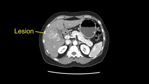
The patient is a 46 year old woman with Hepatitis B. On surveillance, she was found to have a 3.5cm lesion hepatocellular carcinoma predominantly in Segment 5 of her liver. There is no evidence of cirrhosis. The plan is to perform a right sided liver resection.
Right Hepatectomy [Case 17]
Imaging Analysis
Look through the patient's CT scan to identify any areas of concern for the necessary procedure.
Surgeon's Impressions
Watch the following videos of the surgeons preoperative plans for the case. Compare their findings with your own analysis.
OR Footage
Summary of surgeons' findings and slowing down moments:
-
Operating Surgeon
Dr. Greig's imaging findings: Formal right hepatectomy
- Preserve main MHV
- Portal dissection: Dissect RPV, Divide RHA at common trunk
- Sg7 vein in parenchymal bridge
- Divide MHV in Sg5
- Dissection of Sg6 vein at IVC
-
Consulting Surgeon
Dr. Wei's imaging findings: Sg5-6 bisectionectomy, en bloc Sg7 wedge
- Tumor proximity to inflow: Dissect Sg5 PV, Sg8 PV
- Preserve Sg6/7 PV
- Assess outflow: RHV, MHV
- Intraoperative ultrasound for Sg8 inflow
- Ensure blood flow to liver
-
Procedure-specific moments:
- Assess liver tissue
- Future liver remnant
- Tumor proximity to vascular, biliary structures
- Any extrahepatic disease
Watch the following videos from the operation to see some of the key moments in the case.
Post-op Debrief
Watch the following videos for the surgeons summaries of the case.
Key points from this case
- Use intraoperative ultrasound to guide and confirm resection decisions at the time of surgery. Particularly when considering a parenchymal-sparing approach.
- Constantly evaluate intrahepatic landmarks to ensure margins are maintained throughout the transection and adjust transection line as needed.
- Parenchymal bridge can be anticipated based on preoperative imaging. Be alert for small branches for careful mobilization off the IVC.
The issues that the surgeons collectively identified preoperatively:
- Tumor proximity to RPV bifurcation
- Assess liver tissue and future remnant
- Preserve MHV
- Portal dissection: inflow and outflow
- Intraoperative ultrasound to confirm anatomy
- Formal right hepatectomy or Sg 5-6 bisectionectomy with Sg7 wedge
The issues were demonstrated in the intraoperative videos. So I’ll make a couple of comments in this postoperative debriefing after this right hemihepatectomy. First of all, the initial assessment was that the liver was indeed non-cirrhotic and that there’s a single tumor in the right lobe of the liver both by palpation and on ultrasound. And the intraoperative staging did demonstrate the relationship between the tumor and the Segment 8 branch of the portal vein, which would require a right hemihepatectomy. So we proceeded. The portal dissection was really quite uneventful.
The liver was a little more adherent to the cava and after the parenchymal bridge had been taken, we then got around the right hepatic vein and because of the stickiness we had to do that from above rather than from below but the right hepatic vein was controlled.
Parenchymal transection began along Cantlie’s line and as we encountered the Segment 5 vein becoming the middle hepatic vein, we reassessed the relationship between the tumor and the transection line and it appeared to me that it was a little too close. So we revised our transection line, moving it more into Segment 4. So the transection line was revised intraoperatively. Then the liver was transected. The bile duct was divided. The transection was completed and otherwise was unremarkable. So those are the major issues that occurred that distinguished this right hemihepatectomy.