Right Hepatectomy (to right anterior sectionectomy) [Case 16]
Introduction
Clinical History
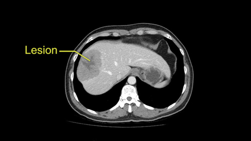
This is a 51 year old man with Hepatitis B cirrhosis with numerous regnerative nodules but stable disease. On ultrasound, a new lesion was found measuring 6.0 x 6.5cm with mild heterogeneous arterial enhancement and possible central necrosis. He has no other liver lesions and no vascular invasion or extrahepatic disease. Liver synthetic function was well preserved. The plan is to perform a right hemihepatectomy.
Right Hepatectomy (to right anterior sectionectomy) [Case 16]
Imaging Analysis
Look through the patient's CT scan to identify any areas of concern for the necessary procedure.
Surgeon's Impressions
Watch the following videos of the surgeons preoperative plans for the case. Compare their findings with your own analysis.
OR Footage
Summary of surgeons' findings and slowing down moments:
Operating surgeon: Dr. Cleary
- Procedure-specific moments:
- Assess liver, disease severity
- Mobilizing the liver
- Dissection at porta hepatis
- Resection decision
- Intrahepatic structures during transection
- Critical imaging findings:
- Relatively healthy liver
- Right hemihepatectomy or anterior sectionectomy
- RHV branches to Sg 7, Sg 8
- Variant portal vein anatomy
Consulting surgeon: Dr. Gallinger
- Procedure-specific moments:
- Parenchymal transection
- Critical imaging findings:
- Variant portal vein bifurcation
- Tumor proximity to anterior RPV branch
- Identify both RHA branches
- Preserve MHV
Watch the following videos from the operation to see some of the key moments in the case.
Post-op Debrief
Watch the following videos for the operating surgeon's summary of the case.
Key points from this case
The issues that the surgeons collectively identified preoperatively:
- Mobilization and assessment of liver
- Variant portal vein anatomy
- Tumor proximity to right portal inflow
- Parenchymal transection and intrahepatic structures
- Tumor proximity to MHV, RHV
- Intraoperative decision on resection
In concluding this patient’s operation, we can reflect back on a number of slowing down moments that occurred throughout the case. The first slowing down moment occurred very early as we opened the abdomen and performed a laparotomy, we found that - first, the liver did not show significant signs of cirrhosis. He had very mild cirrhosis.
The second one was that the tumor was invading through the diaphragm and so therefore it was very important that we identify this before placing our retractors. So what that required was for us to actually resect a segment of the membranous portion of the diaphragm with the tumor en bloc to obtain adequate oncologic clearance and actually prevent the tumor from rupturing during mobilization.
The second slowing down moment occurred during the initial assessment of the liver and the intraoperative ultrasound we performed. The tumor was probably larger than we anticipated and it had more vascular involvement than the preoperative CT scan had indicated. The intraoperative ultrasound indicated that the middle hepatic vein was involved in the tumor but that the right hepatic vein and posterior sectoral inflow was not involved in tumor. We analyzed the volumes of both the left lobe and the right posterior sector and trying to decide between an extended right hepatectomy with middle hepatic vein or an anterior sectionectomy. When we analyzed the volumes, the volume of the left lobe was 32%, and the volume of the posterior sector was 15%. 32% is probably a sufficient future liver remnant for a patient without cirrhosis or without liver disease. We felt that in this patient it might be cutting it a little bit close and so we elected to try and preserve the posterior sector in preserving both the inflow and outflow of the segment as well as a great deal of the parenchyma in that segment. So we elected to perform an anterior sectionectomy and that was a much more difficult operation with a lot more transection than an extended right hepatectomy. But we feel that if we can minimize the risk of post-operative liver failure then that sort of approach is worth it. Certainly we had to take into consideration, the added difficulty of the operation, and possible increased blood loss with a more complex resection, and balance that with the preservation of parenchyma.
Next slowing down moment obviously occurred with the previously identified portal vein anomalies. We were able to identify the right posterior, right anterior, and left portal vein branches independently and we were able to ligate and divide the anterior branch of the right portal vein only. Again, there was no common right portal vein to dissect or identify.
Other slowing down moments occurred throughout the case in really defining the anatomy, as we dissected through Segment 4, this was a relatively straightforward resection. The next slowing down moment was really defining the right posterior portal inflow and then mobilizing along the right hepatic vein because we felt that this was our adequate tumor margin.
The final slowing down moment came in the division of the middle hepatic vein. Since this could not be taken outside of the liver we had to take it inside the liver and this was really the final point of the dissection that allowed us to remove the specimen en bloc.