Choledochectomy [Case 15]
Introduction
Clinical History
A healthy 57-year old woman, had an ultrasound investigation of microscopic hematuria, incidentally revealed a cystic abnormality in her bile duct. Imaging showed a common bile duct dilatation of 19mm. She was asymptomatic with no history of liver disease, jaundice, or choledochalithiasis. The radiologic diagnosis was a Type 1 choledochal cyst. The plan is to do a choledochetomy and choledochojejunostomy reconstruction.
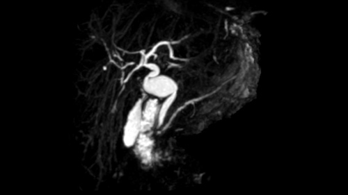
Choledochectomy [Case 15]
Imaging Analysis
Look through the patient's CT scan to identify any areas of concern for the necessary procedure.
Surgeon's Impressions
Watch the following videos of the surgeons preoperative plans for the case. Compare their findings with your own analysis.
OR Footage
Summary of surgeons' findings and slowing down moments:
- Procedure-specific moments:
- Confirm anatomy
- Bile duct margins: proximal and distal
- Preserve GDA, Common hepatic artery
- Visualize portal vein
- Dissection of RHA
- Critical imaging findings:
- Atypical features of choledochal cyst (Type I)
- No evidence of cancer
- No biliary sludge or choledochalithiasis
- Proximity of lesion to RHA
- Length and proximity to common hepatic duct
- Proximity of lesion to pancreatic head
Watch the following videos from the operation to see some of the key moments in the case.
Post-op Debrief
Watch the following videos for the surgeons summaries of the case.
Key points from this case
When anatomy is uncertain, proceed carefully, slowdown and err on the side of caution until you can be certain of the structures.
The issues that the surgeons collectively identified preoperatively:
- Relationship of RHA to common bile duct
- Proximity of cyst to pancreas
- Proximal margin (hepatic duct)
- Distal margin (common bile duct)
So this woman’s choledochectomy went well. There were really no surprises at the time of surgery. She is a thin woman which facilitated the dissection. We could see the cystic dilatation of the bile duct. In our initial dissection around the top of the duodenum, I was a little surprised in that the segment of common bile duct below the cyst was shorter than I expected. The cyst actually went down into the pancreas a little further than I would have anticipated based on the imaging. We dissected out 3 or 4 millimetres of the bile duct out of the pancreas to get well below the cyst. Not technically challenging but it was a bit of a surprise.
At the top end, the hepatic artery was particularly favorable. Once we drop the gallbladder down, and dissected to the right of the common hepatic duct, the right hepatic artery was very evident and we’re able to dissect the common hepatic duct off the right hepatic artery, roll everything off the artery itself. It was a very high lying artery which is very favourable and we were nowhere near the gastroduodenal artery which was well over to the side.
In coming across the neck, there was a second longitudinal structure to the left of what was obviously the common hepatic duct or what I thought was the common hepatic duct. I wasn’t sure whether this was the bifurcation of the bile duct into left and right, or whether its just a nerve or another tubular structure. So we kept that intact and when we divided the neck or just above the neck of the cyst itself, had a good look inside the common hepatic duct and probed it carefully. We could determine that the bifurcation, in fact, was 3 or 4 - 5 or 6 millimeters- actually upstream. That was just a nerve all along the side. There was certainly no additional lumen up the right hand side. So the issue was that during the surgery we were a little surprised to find an additional longitudinal structure that might have been a small bile duct. We preserved it in case if it was a bile duct, we’d include it in the anastomosis but as it turns out, we determined that indeed it was nothing. It was just a nerve, lymphatic structures and tissue running along the side of the bile duct itself. The reconstruction was uneventful with the choledochojejunostomy.