Laparoscopic distal pancreatectomy with possible splenectomy [Case 14]
Introduction
Clinical History
Clinical History
This is a 48-year old woman with history of kidney stones who was referred with an incidental finding of a lesion in the body of the pancreas. The multiseptated cystic lesion measures 3.2 x 3.9 x 4.3cm with an area of gadolinium-enhanced septal thickness. She was asymptomatic with no history of pancreatitis. There is no biliary dilatation and the spleen appeared normal. The plan is to do a laparoscopic distal pancreatectomy with possible splenectomy.
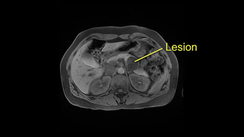
Laparoscopic distal pancreatectomy with possible splenectomy [Case 14]
Imaging Analysis
Look through the patient's CT scan to identify any areas of concern for the necessary procedure.
Laparoscopic distal pancreatectomy with possible splenectomy [Case 14]
Surgeon's Impressions
OR Footage
Summary of surgeons' findings and slowing down moments:
Operating surgeon
- Dr. Cleary's Procedure-specific moments:
- Mobilization of pancreas
- Possible splenic preservation
- Dissection of splenic vessels
- Division at neck of pancreas
- Dissection of pancreatic tail at splenic hilum
- Critical imaging findings:
- Cystic nature of lesion
- Tumor proximity to splenic vein
- Tumor proximity to left adrenal gland
- Posterior pancreas margin: Gerota's fascia
- Anterior pancreas margin: posterior wall of stomach
Consulting surgeon
- Dr. Moulton's procedure-specific moments:
- Tumor proximity to portal vein, tunnel behind neck of pancreas
- Tumor proximity to splenic vein and splenic artery
- Possible splenic preservation
- Retroperitoneal dissection
- Patient imaging findings:
- No formal tunnel dissection required
- Normal ligation of splenic vein and splenic artery
- Warshaw technique for splenic preservation
- Tumor proximity to left adrenal gland
Watch the following videos from the operation to see some of the key moments in the case.
Post-op Debrief
Watch the following videos for the surgeons summaries of the case.
Key points from this case
When deciding whether to preserve the splenic vessels in a spleen preserving distal pancreatectomy, evaluate the anatomical relationship of the vessels at both the transection margin and the tail of the pancreas, and take care in any dissection at the splenic hilum. When performing a Warshaw technique, ensure there is adequate splenic perfusion from short gastric vessels.
The issues that the surgeons collectively identified preoperatively:
- Mobilization of cystic lesion in pancreas
- Dissection of splenic vessels
- Dissection of pancreatic neck
- Proximity of pancreatic tail to spleen
- Decision for splenic preservation
- Tumor proximity to left adrenal gland
- Pancreatic dissection margins
The issues were demonstrated in the intraoperative videos. So in summary, we were successful in performing a spleen preserving distal pancreatectomy laparoscopically in this patient. A few post-operative considerations to reflect upon is the fact that we did a vessel sacrificing procedure or Warshaw procedure. Due to the proximity of the lesion [off] to the splenic vein, I did not think that vessel preservation was appropriate and therefore, we divided the splenic artery and splenic vein proximally in the neck and distally in the splenic hilum and left the spleen on the short gastric vessels. We were able to preserve the gastroepiploic vessels as well as the short gastrics and we felt that splenic perfusion was adequate at the end.
In terms of other post-operative factors, the plane posterior to the pancreas was relatively well preserved and therefore we did not need to enter into Gerota’s fascia or resect the adrenal gland. The lesion itself though was quite adherent to the transverse mesocolon and therefore we did need to resect an avascular portion of transverse mesocolon. This created a mesenteric defect that needed to be closed at the end of the case to reduce the risk of internal hernia in the future.