Right hemihepatectomy (to posterior sectionectomy) [Case 13]
Introduction
Clinical History
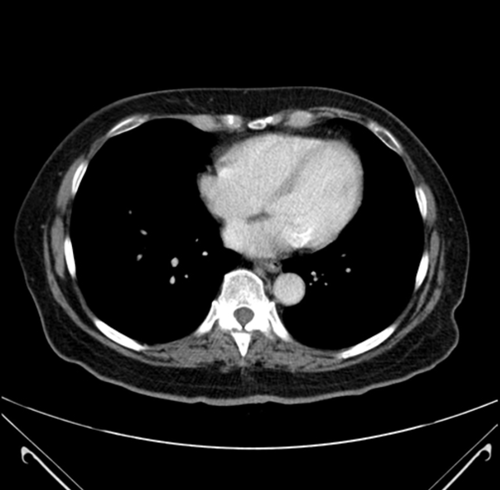
This is a 65 year old woman who had suspected colorectal liver metastases with lesions in: Segment 5 (0.9cm), Segment 6 (1.4cm), and Segment 7 (2.0cm). She underwent a right hemicolectomy and cholecystectomy nine months ago.
On follow-up, the liver lesions were found to be growing in size, with a new lesion in Segment 4a-8 (0.6cm), and intraductal biliary dilatation in Segments 6-7. She underwent chemotherapy and responded well. The lesions decreased in size: Segment 5 (0.8cm), Segment 6 (0.9cm), Segment 7 (0.3cm); with improvement in previously dilated biliary ducts thus suggesting intrabiliary metastatic disease. The plan is to perform a right hemihepatectomy.
Right hemihepatectomy (to posterior sectionectomy) [Case 13]
Imaging Analysis
Look through the patient's CT scan to identify any areas of concern for the necessary procedure.
Right hemihepatectomy (to posterior sectionectomy) [Case 13]
Surgeon's Impressions
OR Footage
Summary of surgeons' findings and slowing down moments:
Procedure-specific moments
- Laparotomy for extrahepatic disease
- Mobilization of right liver
- Intraoperative ultrasound
- Check for liver disease
- Dissection of inflow vessels
- Dissection of PV bifurcation
- Dissection of outflow vessels
- Avoid common bile duct
- Right hepatic vein dissection
- Hanging maneuvre
- Pringle maneuvre
Patient-specific moments
- Review tumor locations on previous imaging
- Proximity of tumors to vascular structures
- Intrahepatic ductal dilatation in Sg 7
- Sg 7 branch at right portal vein bifurcation
- Divide bile duct in liver
- Possible posterior sectorectomy, local resection
- Biliary guide or contrast
- Inspect lymph nodes at hepatoduodenal ligament
Watch the following videos from the operation to see some of the key moments in the case.
Post-op Debrief
Watch the following videos for the surgeons summaries of the case.
Key points from this case
Careful review of preoperative imaging is required particularly for previously identified tumors that have decreased in size following chemotherapy.
When revising surgical options, consider future liver remnant and implications for possible recurrence and resection.
The issues that the surgeons collectively identified preoperatively:
- Sg 6-7 intrahepatic ductal dilatation
- Dissection of hepatic outflow vessels
- Dissection of hepatic inflow vessels
- Proximity of tumors to vascular structures
- Dissection of common bile duct
- Determining liver transection plane
- Mobilization of right liver
- Multiple small size tumors
So based on the findings that we found at the time of surgery, we did modify the plan and we had some additional intraoperative decision making moments. Importantly, one of the tumors, particularly the one the most medial in the liver, had an excellent response to chemotherapy.
The tumor is Segment 4a/8 was actually not visible. At the time of intraoperative ultrasound, we decided to modify the planned procedure. Mostly to preserve as much parenchyma of the liver as feasible. In addition, to preserve as much inflow and outflow of the liver so if her disease were to recur, we would have greater options in terms of further re-resection at a later date.
So based on this I did not feel that the patient intraoperatively needed a complete right hemihepatectomy. That she did not have to have all of Segment 5-8 removed and we elected to perform a posterior sectionectomy, which is a Segment 6-7 resection encompassing a small portion of Segment 5 to remove the small tumor that was still present.
The reason I felt a Segment 6-7 was needed in its entirety was that we could see again the intraductal dilatation in Segment 7 at intraoperative ultrasound. In fact, the dilatation was slightly worse than it was preoperatively so we were highly suspicious of a tumor high in Segment 7 that could not be removed with a simple wedge resection.
There were several slowing down moments. Always when you modify the planned procedure, I always take a very careful look at the entire CT scan again instead of just going ahead without reviewing it because there may be small important questions that you want to understand about the anatomy before you proceed. With a posterior sectionectomy, you need to take the inflow of the blood vessels deeper in the liver in a more selective way. At the time of surgery, we re-reviewed the CT scan and we could see that the anatomy for the 6-7 and 5-8 portal system and the arterial system was extrahepatic, which made this maneuver and this operation quite straightforward.
The Segment 6-7 hepatic artery, 6-7 and 5-8 portal branches were clearly separated of each other and clearly outside the liver so we could be very confident that we identified at the time of surgery the same thing we could see on the CT scan. We also confirmed the presence of the left portal system before we stapled and divided the inflow, the Segment 6-7 portal vein at the level of the hepatic artery, extrahepatically.
We also discussed whether we should or can take the right hepatic vein which we’re preserving part of the right liver we are now leaving in, would be adequately drained. And we did not take the right hepatic vein as previously planned. So those are the decisions that we made intraoperatively, the slowing down moments that made us decide whether or not a posterior sectionectomy was an appropriate choice in this patient.