Mid-bile duct resection [Case 10]
Introduction
A 70-year old man had an incidental finding of suspected cholangiocarcinoma. He was asymptomatic but has a distended colon from other comorbidities.
The CT scan revealed a mass in the biliary tract and intrahepatic duct dilatation.
The plan is to do a mid bile duct resection with cholecystectomy and biliary reconstruction.
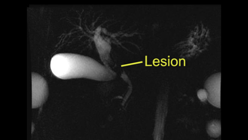
Mid-bile duct resection [Case 10]
Imaging Analysis
Look through the patient's CT scan to identify any areas of concern for the necessary procedure.
Mild-bile duct resection [Case 10]
Surgeon's Impressions
OR Footage
Summary of surgeons' findings and slowing down moments:
Operating surgeon
- Dr. Moulton's critical imaging findings:
- Proximal bile duct margin
- Distal bile duct margin
- Tumor proximity to right hepatic artery
Consulting surgeon
- Dr. Greig's critical imaging findings:
- Proximal biliary tract margin: common hepatic duct
- Distal biliary tract margin: common bile duct
- Intraoperative decision for Whipple operation
- Tumor proximity to right hepatic artery
Watch the following videos from the operation to see some of the key moments in the case.
Watch the following video for the operating surgeon's summary of the case.
Operating surgeon: Dr. Carol-anne Moulton
The issues were demonstrated in the intraoperative videos. So we’ve completed the bile duct resection and I think judging by how it felt, it appeared to be a cholangiocarcinoma. Just by palpation. It didn’t appear to be involving the gallbladder and the bile duct was soft above the tumor, and soft below the tumor above the pancreas. So I was very happy that we got adequate margins. The frozen sections of both the upper limit and the lower limit were negative.
We made a decision to come across the bile duct right at the bifurcation of the right and the left and to sew to one duct only and then reconstruct it with a Roux-en-Y hepaticojejunostomy. There were no surprising features in this case, other than a very large colon which presented more as a nuisance to try to keep out of the way. That was because of his pre-op co-morbidities of a spastic paresis. We will await the formal pathology but I’m pretty sure this was a cholangiocarcinoma of the mid bile duct, which is actually quite uncommon. We were able to do a mid bile duct resection with a lymphadenectomy.
Consulting Surgeon: Dr. Mariano Gimenez
Transcripción en Español
Bueno, tanto ustedes como nosotros estuvimos viendo los cuatro pasos de la cirugía realizada de resección. El primer paso que hace el cirujano es buscar, nódulos alejados con la idea de que el patólogo le diga si hay enfermedad diseminada. En este caso es realmente muy poco probable, porque es un tumor muy insipiente por suerte, pero es una buena técnica para en ese momento, si hubiera tumor en los nódulos hepato-aorticos, se hablaría de una enfermedad diseminada y no tendría sentido hacer la resección.
Después el cirujano empieza a hacer maniobras, que son reversibles. Y esto es importante que ustedes lo tengan presente. El cirujano empieza a ver la relación de la arteria con el tumor, para confirmar lo que le mostraron las imágenes preoperatorias y ahí observa que tanto la arteria hepática común, como la arteria hepática derecha, están separadas, se pueden separar relativamente fácilmente con habilidad como lo mostro el cirujano, las pudo separar de la vía biliar y después fue a ver la porta y la relación de la porta con el tumor y también vio que que podía separa los planos.
Una vez que hace eso, hace una resección de la vesícula, y la hace retrograda con la idea de sacar la pieza en bloc, eso es algo muy importante, sacar la pieza en bloque. No cortando de a pedazos sino sacar todo el bloque junto. Busca también, nódulos retro pórtales y una vez que corta la vía biliar por debajo, lo hizo con una stapler. Uno podría hacerlo con una ligadura simple. El stapler es mas rápido, pero más caro, con una ligadura simple es mas económico, y los resultados son iguales. Pero es importante que hacerlo con una ligadura, ósea cerrando esto, para que no se disemine en el campo operatorio, bilis que estuvo en relación con el tumor; y esto es importante. No es cortar si no que es ligar, con stapler o con ligaduras y ahí cortar. Una vez que hizo esto, ya se vuelve irreversible el procedimiento y empieza a hacer la resección de la vía biliar y todo los nódulos linfáticos, y la grasa que esta alrededor de la vía biliar, dejando pelada la arteria hepática y la vena porta. Va hacia arriba, que ya la había reparado previamente y hace la resección y después como habíamos hablado en el preparatorio, hace la hepático-jejuno anastomosis.
En definitiva, se pudo hacer en la cirugía lo que habíamos charlado y habíamos planteado en el preparatorio. No hubo que hacer otras maniobras por suerte. porque es una cirugía mucho más sencilla y la reconstrucción en Y-de-roux con asa de intestino es la norma y es lo que hizo el cirujano en forma brillante.
Consulting Surgeon: Dr. Mariano Gimenez
English Translation
Okay, we were watching the four main steps of the surgery. The first step was to assess the hepatic artery lymph node with the idea of asking the pathologist if there is any spread of disease. In this case, it is very unlikely because this is a very early stage tumor. However, it is a good technique if there was a tumor in the hepatic aortic nodules, because we would be in the presence of disseminated disease and there will be no advantage in performing a resection.
Afterwards, the surgeon starts to perform maneuvers that are not irreversible; please keep this in mind. The surgeon starts to check the relation of the artery and the tumor, to confirm what was shown by the pre-operative images, and how far apart the common hepatic artery and the right hepatic artery are. These arteries can be separated fairly easy as it was shown by the surgeon. They separated the arteries from the bile duct, and then assessed the relation of the portal vein with the tumor which was also easily dissected.
Once this was done, a resection of the gallbladder was performed, and this was done in a top-down fashion with the idea of performing an en bloc resection. It is really important to resect the entire specimen en bloc rather than in separate pieces. The surgeon also looks for retro-portals nodes, and then they cut the bile duct distally with a stapler. Another option is to cut the bile duct between simple ties. The stapler is faster, and more expensive; whereas the simple suture is cheaper and the results are the same. But it is important to close the duct, so there is no bile leakage in the area of the surgery. This is important, you shouldn’t cut first, you need to close it first with a stapler or the suture, and then cut. Once this was completed, the process is irreversible; then they follow with the resection of the bile duct with all the lymphatic nodes, and all the tissue that is around the bile duct, leaving the hepatic artery and the portal vein clear and intact. Then they complete the resection as we discussed during the pre-op analysis, and perform a hepaticojejunal anastomosis.
In summary, it was possible to perform the surgery that we have discussed and proposed during the pre-op assessment. Luckily, there was no need for other maneuvers, so it was a fairly simple surgery and the surgeon used a Roux-en-Y reconstruction, and it was done brilliantly by the surgeon.